
Welcome to the Laboratory of Neural Crest and Enteric Nervous System Development
Studying how cells transform from one cell type into another since 2017
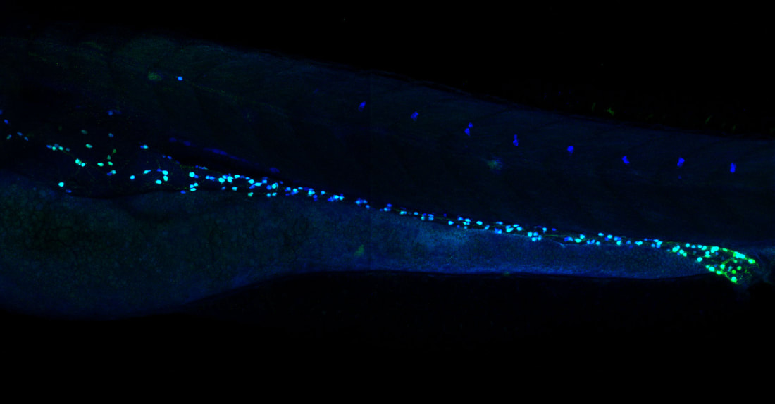
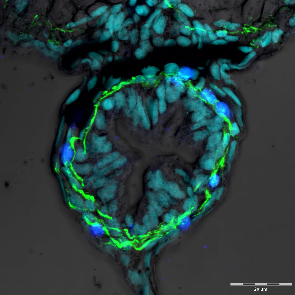
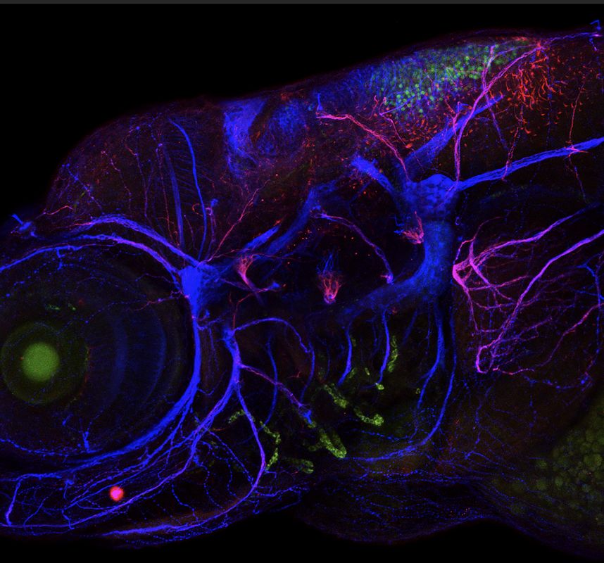
We study how genetic, cellular and environmental factors contribute to and influence stem cell and nervous system development in vivo. Our mission is to use a systems-level approach to enhance understanding of fundamental mechanisms that orchestrate stem cell transformation into specialized cells and tissues. Our approach integrates and leverages zebrafish organismal-level, tissue-level and cellular-level developmental biology, synthetic & recombinant gene expression paradigms, single-cell omics, in vivo high resolution microscopy and computational methods.
Our ultimate goal is to increase foundational knowledge to help build better therapies and cures for neurodevelopmental defects and cancer.
For our current research see here.
Studying how cells transform from one cell type into another since 2017
We study how genetic, cellular and environmental factors contribute to and influence stem cell and nervous system development in vivo. Our mission is to use a systems-level approach to enhance understanding of fundamental mechanisms that orchestrate stem cell transformation into specialized cells and tissues. Our approach integrates and leverages zebrafish organismal-level, tissue-level and cellular-level developmental biology, synthetic & recombinant gene expression paradigms, single-cell omics, in vivo high resolution microscopy and computational methods.
Our ultimate goal is to increase foundational knowledge to help build better therapies and cures for neurodevelopmental defects and cancer.
For our current research see here.
Lab logo design by James Tallman.
Dr. Uribe recognizes prior research support from foundations and institutes that funded scientific training, including NIH and The Burroughs Wellcome Fund, via NRSA and PDEP awards, respectively.
The Laboratory of Neural Crest and Enteric Nervous System Development acknowledges funding from multiple sources, including the NIH, NSF, CPRIT and John S. Dunn Foundation.

|
|